
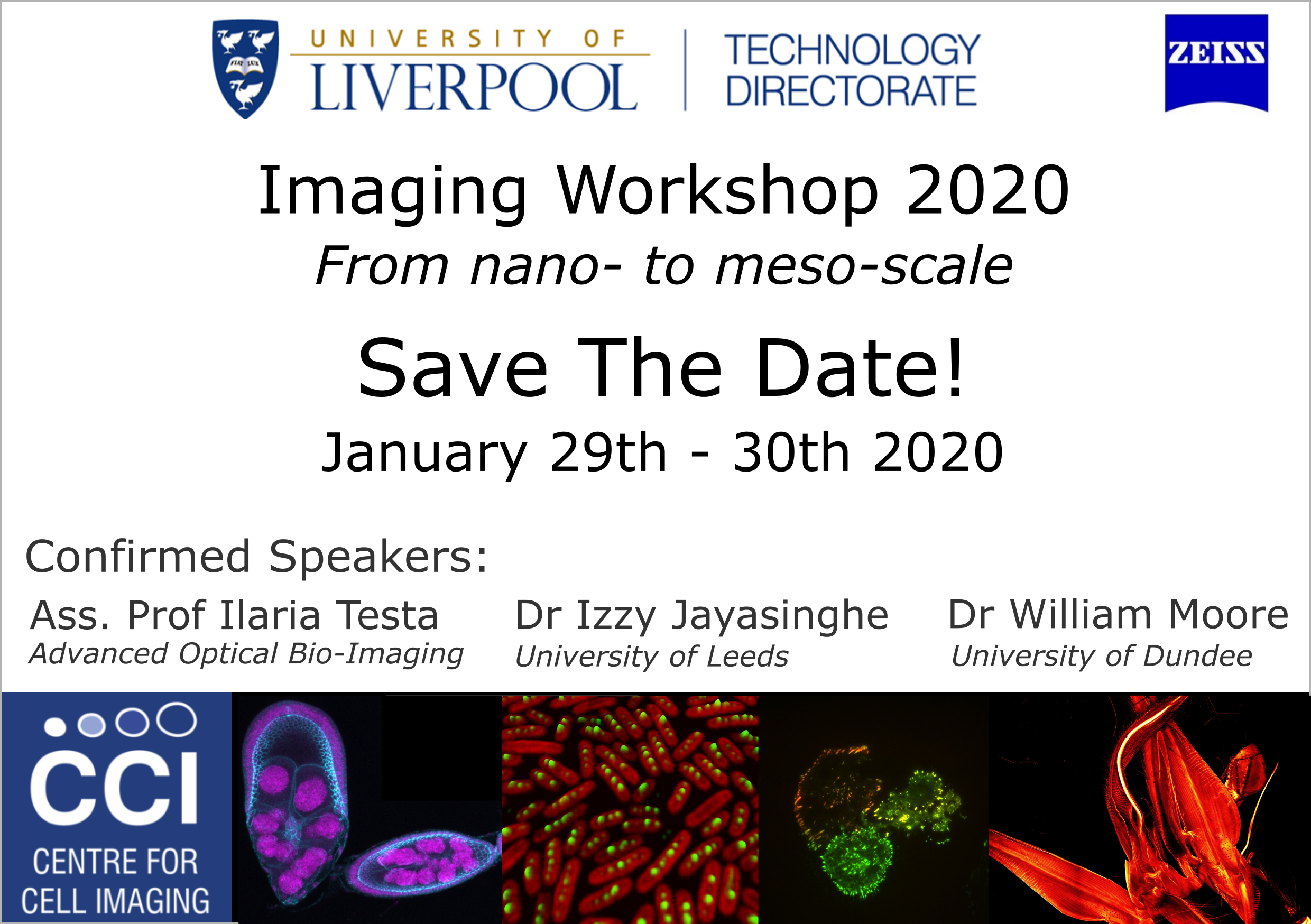
2020 Imaging Workshop: From nanoscale to intravital imaging
We are pleased to announce that the next CCI imaging workshop will take place on 29th and 30th January 2020. This two day workshop aims to provide existing and potential users of the facility with a better idea of the services we offer, the work that is conducted here and some of the cutting edge science happening in the Imaging field. The first day is in symposium style and the second will have a focus on facility tours and image analysis with an IMARIS workshop.
You can download the flyer for the event here.
We (and others) will be tweeting leading up to the event and in real time with the hashtag #CCIwkshp. We do however ask that attendees respect work tagged with the "No Sharing Please" symbol.
Date & Location
The imaging workshop was held on the 29th and 30th January 2020. The main seminars were in the LMI Conference Centre. The Image Analysis hands-on sessions was in a University of Liverpool PC teaching room.
Program
Day 1: Symposium
- 9:30-9:40: Introduction - Violaine Sée & Daimark Bennett (CCI), Tobias Zech (Biomedical Imaging Facility)
- 9:40-10:00: Foundation in microscopy - Tom Waring (University of Liverpool)
- 10:00-10:40: Microscopy techniques evolution, a historical and technical perspective - Chris Power (Carl Zeiss UK)
- 10:40-11:00: Advanced techniques in microscopy starting with F… : FRAP and FRET - Nicolas Sergent (Carl Zeiss UK)
- 11:00-11:25: Coffee break
- 11:25-11:50: Fast Imaging - Geraint Wilde (Andor)
- 11:50-12:50: Selected short talks from imaging scientists in Liverpool.
- 12:50-13:45: Lunch
- 13:45-14:25: Keynote #1: Advancing super-resolution analyses of protein clustering with DNA-PAINT and ExM - Izzy Jayasinghe (University of Leeds)
- 14:25-15:05: Keynote #2: Live cell imaging at the nanoscale - Francesca Pennacchietti (Science for Life Laboratory, Karolinska Institute, Stockholm, Sweden)
- 15:05-15:25: Coffee break
- 15:25-16:05: Keynote #3: Unraveling quantitative dynamics of mammary tissue and tumors using (intravital) microscopy - Colinda Sheele (Netherland Cancer Institute, Amsterdam)
- 16:05-16:30: Using OMERO for imaging workflows and publication - William Moore (University of Dundee, UK)
- 16:30-16:55: Introduction to Imaris - Anna Paszulewicz (Imaris, Oxford Instruments)
- 16:55-17:00 Concluding remarks and best short talk prize
- 17:00: Tours of imaging facilities
Day 2: Imaris Image Analysis Training Course
Day 2 - Anna Paszulewicz, M.Sc.; Global Product Specialist - Imaris
- 09:00-11:00 Imaris for beginners - get started with Imaris, 3D visualization and image segmentation, time-lapse analysis
- 11:00-11:20: Coffee break
- 11:20-16:30 Imaris for Intermediate users - clean your data-set, enhance volume visualisation, improved segmentation, object tracking inside a moving object, classifying objects, animations...
Registration
Registration is not yet open.
Registration is now closed for the 2020 CCI Imaging Workshop. Thank you to everyone who has registered, we look forward to seeing you on the 29th January.
Short Talks
The following short talks were selected:
- Lorna Young (ITM): Beta-pix and Myosin-18a are essential for adhesion-nucleus coupling required for breast cancer cell invasion
- Hammed Badmos (IIB): Ataxin-7 is required for the maintenance of Drosophila egg chamber epithelial polarity
- Manohar Prasad Koduri (School of Engineering): Novel fluorescent based Nano Oxy-PH sensors for 3D Tissue engineering applications
- Mengru Yang (IIB): Biogenesis of bacteria microcompartments
- Anne Reversat (ITM): Leukocyte migration in 3D environments
Congratulations to Anne Reversat (Tobias Zech Group, ITM) who won the best short-talk prize as judged by the invited speakers.
Invited Speakers
Izzy Jayasinghe:
Izzy is a lecturer and a UKRI Future Leader Fellow, currently leading the Nanoscale Microscopy Group in the University of Leeds. She is interested in how the spatial organisation of signalling proteins and heterogeneity in their regulation can enable locally regulated intracellular second messenger systems. To understand these features, Izzy works on developing novel super-resolution imaging and analysis protocols, new imaging chemistries and antibody-conjugated markers. Her upcoming fellowship specifically explores avenues to miniaturise super-resolution microscopy.
Francesca Pennacchietti:
Francesca is a postdoctoral researcher working with Ass. Prof. Ilaria Testa at the Science for Life Laboratory at the Royal Institute of Technology (KTH, Stockholm, Sweden). The goal of the Testa lab is to develop the novel paradigms made available by super-resolution microscopy to address contemporary challenges in biophysics and neuroscience. Their work pushes forward the quantitative aspect of live cell imaging by setting-up and applying different concepts of super-resolution microscopy based on target switching such as automated STED microscopy.
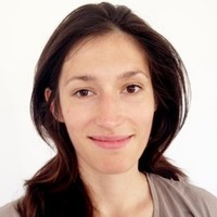
Colinda Scheele:
Colinda is a PhD student at the Netherlands Cancer Institute in the lab of Jacco van Rheenen. The aim of her work is to understand how the healthy mammary gland develops and grows and how these processes are perturbed during mammary tumor development. For this she uses whole organ 3D imaging techniques, as well as intravital microscopy.
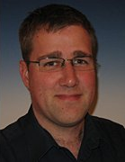
William Moore:
William is a software developer on the OME project, based in Jason Swedlow's lab at the University of Dundee. William’s primary focus is on the OMERO.web client and other OMERO web-based apps such as OMERO.figure and OMERO.iviewer.
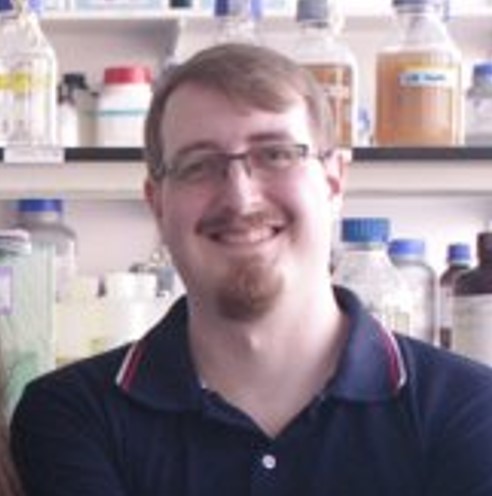
Thomas Waring:
Tom is a PhD student in the Institute of Translational Medicine at the University of Liverpool in the lab of Dr Tobias Zech. His research focuses on assessing mechanotransduction using microscopy-based techniques such as FRET tension biosensors. In 2019, Tom provided valuable support to the Centre for Cell Imaging whilst working his 3 month internship in the imaging facility.
Anna Paszulewicz:
Anna is a Global Product Specialist with Imaris. Anna graduated in Biophysics with a strong emphasis on fluorescence microscopy techniques and image analysis from Jagiellonian University in Krakow. Her professional career has been connected to Andor and Bitplane where she has enjoyed the pleasure of teaching Imaris and creating new image analysis workflows for life scientists.
Chris Power:
Chris is a 3D Imaging Specialist at Carl Zeiss UK. As project manager he has overseen the development of the Zeiss Lightsheet Z1 from optical bench to market and continues to support users of this and other technologies.
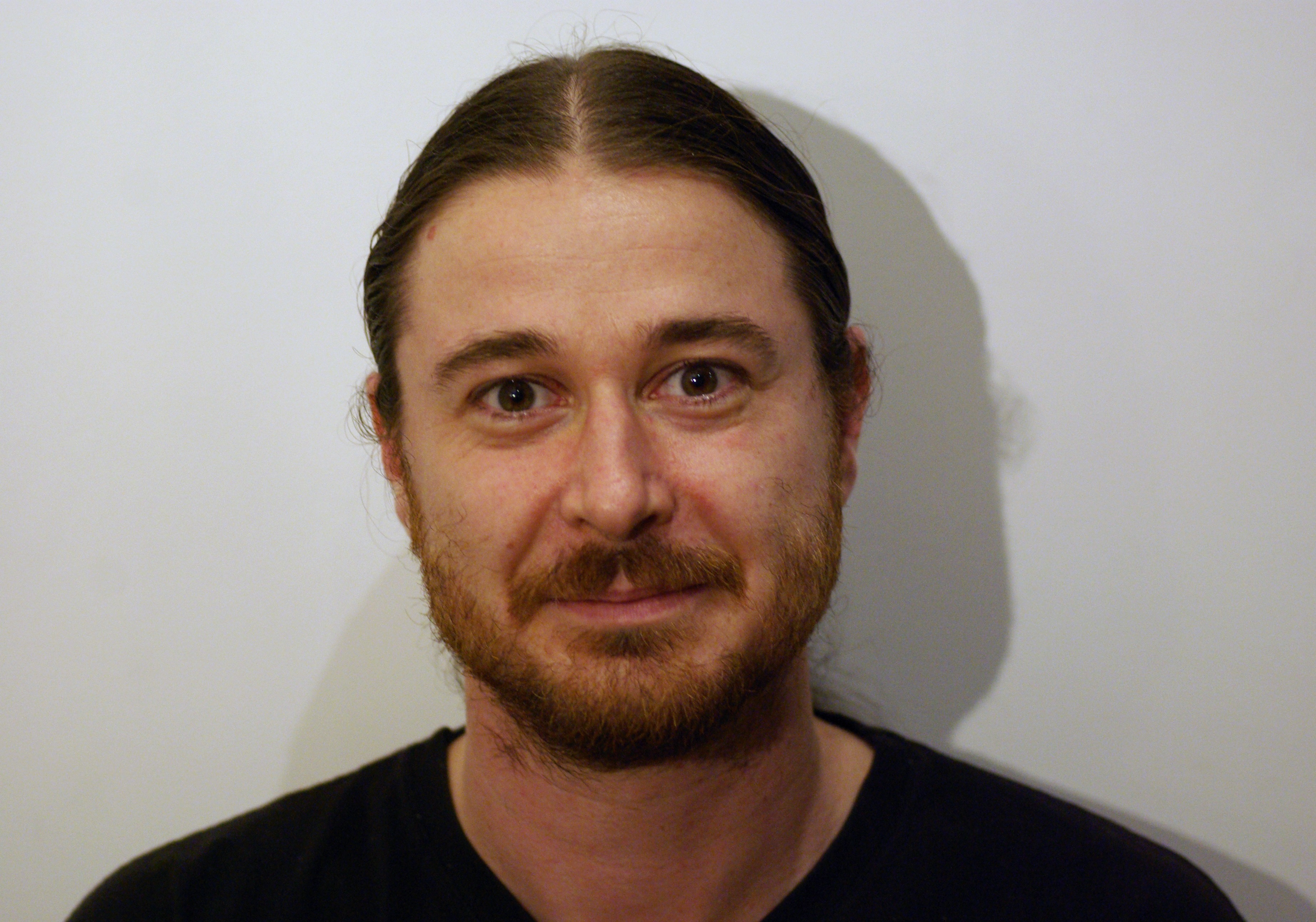
Nicolas Sergent:
Nick joined Zeiss in 2010 having completed a PhD in Physics at Kings College London working in Phosphorescence and Fluorescence Life Time Imaging. He initially joining the service team as a field service engineer and then technical support engineer, supporting confocal, multiphoton and super-resolution (SIM and PALM/STORM) systems in the UK. In 2014 he joined the Microscopy Application team, supporting customers and the UK sales team for 3D Products (Confocal, Multi-photon, TIRF, Super-resolution) in Life Sciences.
Geraint Wilde:
Geraint attained a Ph.D. in Neuroscience in 1997 from the University of Southampton, UK, after which he continued in his interest in neuroscience with a postdoctoral position at the University of Warwick, UK. Having developed an interest in microscopy, he moved to the University of Liverpool, UK, to work in the laboratory of Michael White, focusing on intercellular signaling and gene expression through live-cell imaging. Dr. Wilde eventually pursued a commercial career in microscopy. He joined Andor Technology in 2009, where he is now product manager for microscopy solutions.
Sponsors
The CCI symposium is free to attend thanks to the generous sponsorship of commercial and industrial partners. If you are interested in sponsoring the workshop or would like more details, please get in touch.
The CCI wishes to thank the following companies for sponsoring the Workshop (click for links):
© Liverpool Centre for Cell Imaging