
Photothermal
Stand:
Nikon Eclipse Ti-E
Light source:
- 523
- 633
Polychromatic Mercury Arc Lamp
Detector:
The photothermal microscope principally uses a photodiode detection system, however a high-speed Andor Neo sCMOS or high-sensitivity Hamamatsu EM-CCD camera can be installed for fluorescent imaging.
Application Notes:
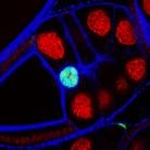
Flurorescent proteins and dyes are great labels which can be used alone or conjugated to antibodies and proteins to demark structures or compartments within a cell. The use of gold nanoparticles has historically been limited to electron microscopy, as these typically cannot be imaged with visible light.
Photothermal microscopy also called LISNa (Light-Induced Scattering around a Nanoabsorber) works by probing the laser scattering of nanoparticles in a sample. This allows for the imaging of GNPs in live or fixed samples. Furthermore the technique can be combined with transmitted light microscopy and widefield fluorescence on the same stand.
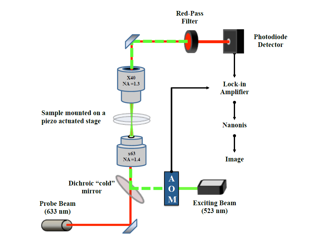
Schematic of a typical photothermal setup. From the thesis of Dan Nieves (available here).
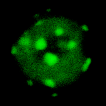
This technique is highly suited to a range of applications involving metal nanoparticles, such as single-molecule tracking and visualiasation of GNP-loaded compartments.
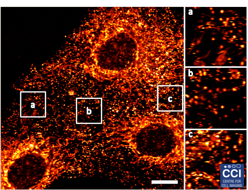
Above, fibroblast growth factor 2 (FGF2) was labelled with gold nanoparticles. These data (courtesy of Daniel Nieves formerly of the Lévy Lab) help to explain how FGF2 interacts with ligands on the cell surface.
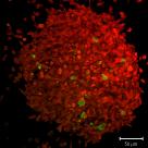
Given the nature of the technique, PT microscopy can also be used for the label-free imaging of red blood cells in tissue sections (below shows a 5μm section of a mouse liver).
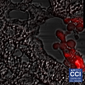
The sample above was provided courtesy of Lauren Scarfe (Murray Lab). Imaging and post-processing by Dave Mason (CCI).
Please note that this system is only available to facility users through collaboration with Raphaël Lévy (rapREMOVEMEha@REMOVEMEliveREMOVEMErpoolREMOVEME.aREMOVEMEc.REMOVEMEuk) who should be contacted directly for more information.
© Liverpool Centre for Cell Imaging