
Incucyte S3
Stand:
S3
LED:
- 488
- 561
Objectives:
Other Features:
Application Notes:
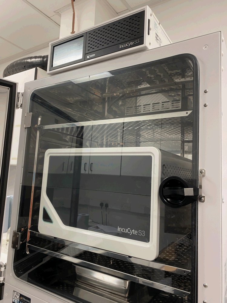
IncyCyte is an incubator microscope system for live cell imaging. The system includes a microscope with enhanced phase contrast and two-color (green/red) fluorescence channels. The system resides inside a standard cell culture incubator and contains a microscope controller, a 30 TB data server and Incucyte image analysis software.

The system allows time-lapse, live cell imaging of cells in a variety of vessels, including 30 mm dishes, tissue culture plates, cell culture flasks and multi-well chambers. Images can be reviewed and analyzed from a networked remote computer without opening the incubator or disturbing the cells. The system is especially suitable for monitoring cell lines, cell growth and transgene expression, and for assay development. The analysis software suite includes validated assays for cell migration, apoptosis, cell proliferation, angiogenesis, reporter gene expression and more.
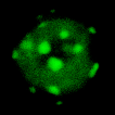
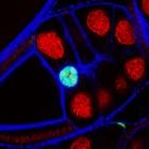
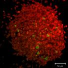
Above, live cells were imaged over ~ two days. Data courtesy of Milan Collins (Zech lab).
Please refer to the following table for the resolution of the images created with the respective objectives.
| Objective | Resolution |
|---|---|
| 4× | 2.82 µm/px |
| 10× | 1.24 µm/px |
| 20× | 0.62 µm/px |
Publication Reference:
This piece of equipment was funded by INSERT RELEVANT INFORMATION IF APPLICABLE. Please cite this grant code when publishing work conducted using this hardware.
© Liverpool Centre for Cell Imaging