
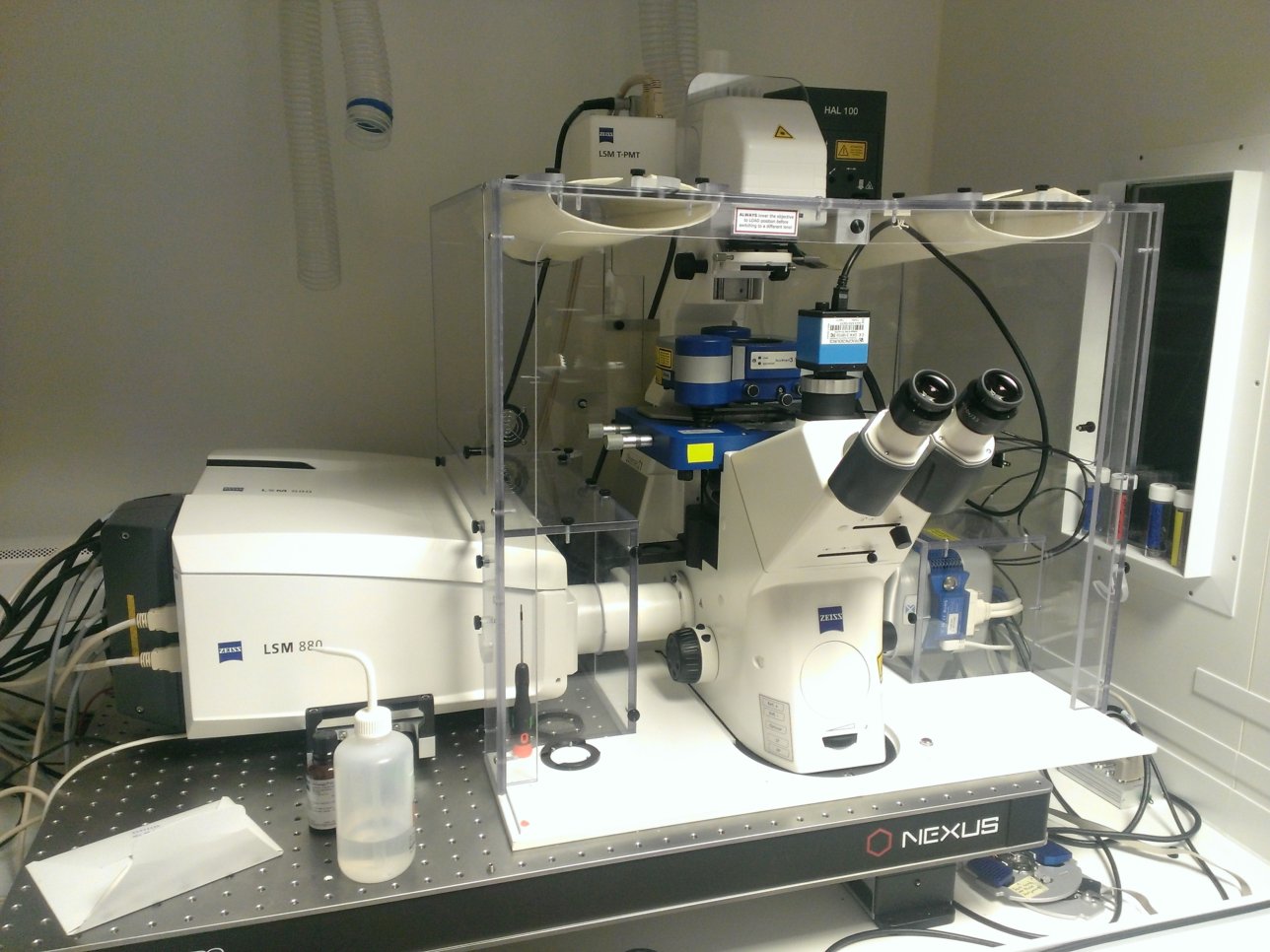
880 BioAFM
Stand:
Zeiss Axio Observer Z1
Laser Lines:
- 405
- 458
- 488
- 514
- 561
- 633
Objectives:
Other Features:
Application Notes:
The 880 BioAFM is a cutting-edge combination Laser Scanning Confocal and Atomic Force Microscope. The confocal (built on a Zeiss LSM 880 platform) has very high sensitivity, speed and optical resolution with motorised control in X, Y and Z, spectral detection and environmental control.

Although not a new technology, Atomic Force Microscopy is reinventing itself in combination with optical imaging, making the study of biological systems with AFM more amenable.
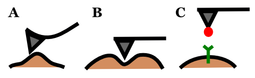
Advanced cantilever and tip design allow the application of AFM to studies of (A) membrane topology, (B) physical perturbation, (C) protein-protein interaction, and many more.

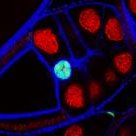
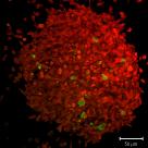
The above cells from a patient suffering from Hutchinson-Gilford Progeria Syndrome were imaged on the microscope, both in atomic force microscope and light microscope mode. The cells were imaged whilst alive. In AFM mode, the topography of the cell was recorded, resulting a height map. Simultaneously, the stiffness of the same region was also recorded and is displayed as a stiffness map. Using an integrated camera, a transmitted light image of the cell was captured and the stiffness map has been overlaid for reference.
Data courtesy of Tom Waring (Zech lab).
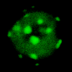
In addition to highly sensitive detectors and AFM functionality, the BioAFM is also equiped for Total Internal Reflection Fluorescence Microscopy (TIR-FM). This technique allows the investigation of surface-localised phenomena (˜100nm from the coverslip) such as membranes or (below) focal adhesions.
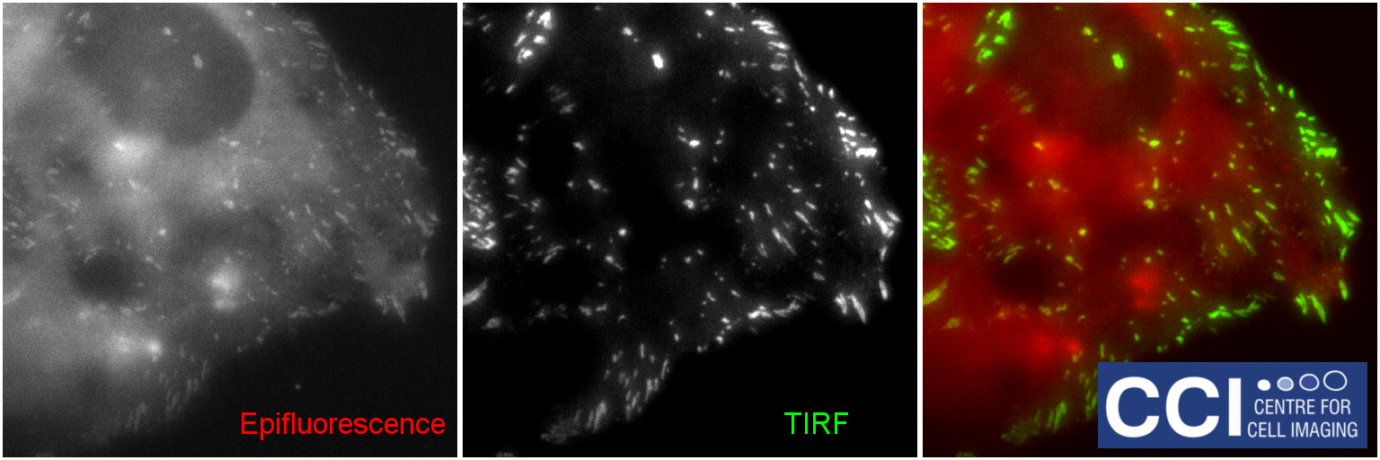
HeLa cells transfected with DLC1-GFP. Data courtesy of Jennifier Francis (Lévy Lab).
Publication Reference:
This piece of equipment was funded by BBSRC grant number BB/M012441/1. Please cite this grant code when publishing work conducted using this hardware.
© Liverpool Centre for Cell Imaging