
LSM 710
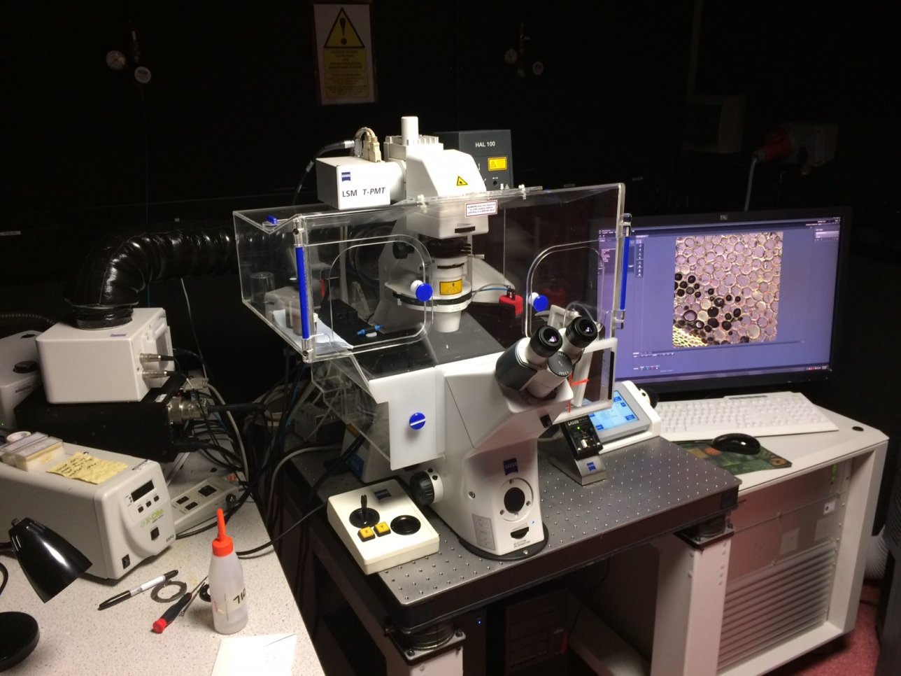
Stand:
Zeiss Axio Observer Z.1
Laser Lines:
- 458
- 488
- 514
- 561
- 633
Objectives:
Other Features:
Application Notes:
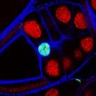
Traditional fluorescent microscopy relies on a single emission filter to select an output range. Spectral detection allows for the acquisition of data across a range of wavelengths, accumulating more of the emitted light.
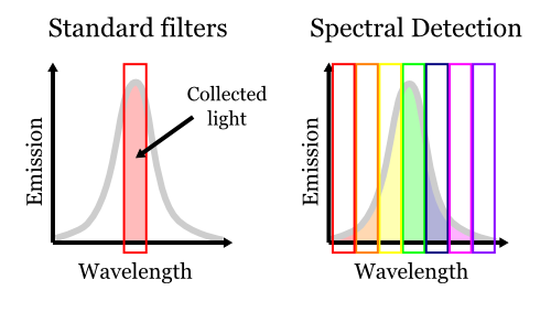
Spectral detection also allows multiple spectra to be 'unmixed' if you know the shape of the individual spectra. This is particularly useful to spatially separate closely related fluorophores (eg. Alexa488 and eGFP).
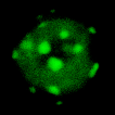
Some samples, particularly those from plant tissues, produce a lot of autofluorescence, which makes it impossible to identify signals from other fluorescent proteins.
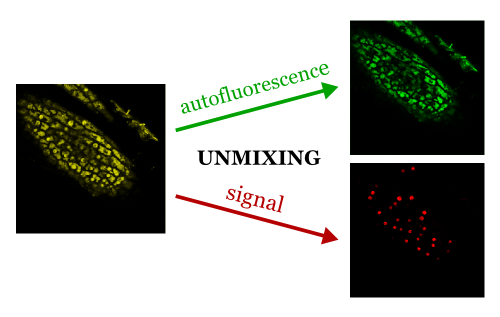
The example above (courtesy of Peter Gould of the Hall Group) shows a leaf of Arabidopsis expressing a red fluorescent protein and imaged in spectral mode. Only after unmixing can the autofluorescence (green) be optically separated from the signal of interest (red).
© Liverpool Centre for Cell Imaging